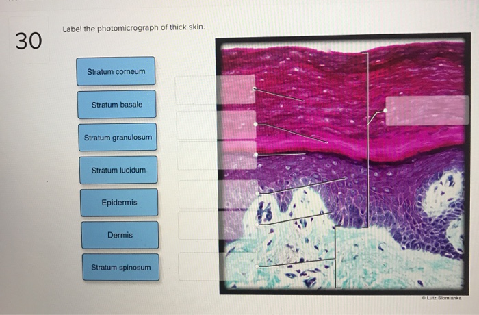
40 label the photomicrograph of thick skin
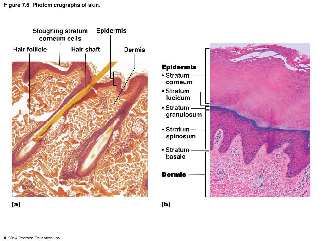
photomicrograph of the epidermal layer in thick skin photomicrograph of the epidermal layer in thick skin Diagram | Quizlet photomicrograph of the epidermal layer in thick skin 5.0 (2 reviews) + − Learn Test Match Created by abba_dabba_17 Terms in this set (6) Term stratum corneum Location Term stratum lucidum Location Term stratum granulosum Location Term stratum spinosum Location Term Figure 7.1: Photomicrograph of Skin Diagram Figure 7.1: Photomicrograph of Skin 3.0 (2 reviews) + − Learn Test Match Created by SophiaVisaggio Terms in this set (5) Term dermal papillae Location Term epidermis Location Term papillary layer of dermis Location Term reticular layer of dermis Location Term hypodermis Location Students also viewed photomicrograph of the epidermal layer in thi…
Lab 9: Pre-Lab Homework Flashcards | Quizlet Label the photomicrograph of thin skin. In general, nerves from the posterior division of the brachial plexus tend to innervate muscles that extend the parts of the upper limb. True/False True Match the label to its appropriate spinal cord component. -dorsal root ganglion -white matter -gray matter -epidural space -dorsal ramus

Label the photomicrograph of thick skin
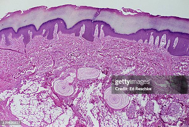
Solved Label the photomicrograph of thick skin. Stratum | Chegg.com The stratum corneum helps to protect the body from damage and dehydration. View the full answer Step 2/2 Final answer Transcribed image text: Label the photomicrograph of thick skin. Stratum corneum Stratum basale Stratum granulosum Stratum lucidum Epidermis Dermis Stratum spinosum Previous question Next question This problem has been solved! Label the photomicrograph of thin skin Label The Photomicrograph Of Thick Skin. Stratum Granulosum Stratum Spinosum Epidermis Stratum Corneum Stratum Basale Dermis Stratum Lucidum ResetZoom Posted one year ago. Q: Label the photomicrograph in Figure 7.4. Examine a slide of hairy skin and identify the structures in Figure 7.4. Solved Chapter Six Iniegumen 19 Label the following | Chegg.com Expert Answer 100% (3 ratings) b. This section of skin is likely from hairless skin of palm or sole which is called thick skin. c. … View the full answer Transcribed image text: Chapter Six Iniegumen 19 Label the following photomicrograph of the skin in part (al, then answer parts tbl and (e) of this question (elow the diagram 10 b.
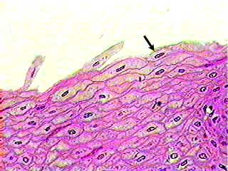
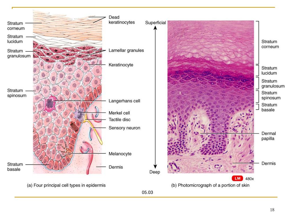
Label the photomicrograph of thick skin. Question: Label the photomicrograph of thin skin Expert Answer. 100% (25 ratings) Transcribed image text: Label the photomicrograph of thin skin. photomicrographs of thin skin Flashcards | Quizlet photomicrographs of thin skin stratum corneum Click the card to flip 👆 Click the card to flip 👆 1 / 4 Flashcards Learn Test Match Created by Madison_Tacquard Terms in this set (4) stratum corneum sebaceous gland hair follicle dense irregular CT of the reticular layer of the dermis Students also viewed LP5 LAB 6 terms jordanmmertens photomicrograph of thick skin Diagram Start studying photomicrograph of thick skin. Learn vocabulary, terms, and more with flashcards, games, and other study tools. hello quizlet Home Subjects Expert solutions Log in Sign up photomicrograph of thick skin Learn Test Match Learn Test Match Created by mckennawebber Terms in this set (7) Term epidermis(stratum corneum - stratum basale) Anatomy and Physiology Homework Chapter 6 Flashcards The stratum lucidum is a thin zone superficial to the stratum granulosum, seen only in thick skin. The stratum corneum consists of up to 30 layers of dead, scaly, keratinized cells that form a durable surface layer. Label the cell types found in the skin. -Exfoliating keratinocytes -Dead keratinocytes -Living keratinocytes -Dendritic cell
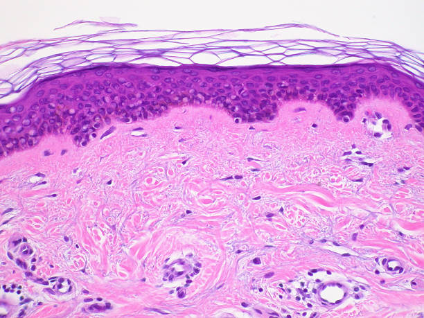
Question: Label the photomicrograph of thick skin Label the photomicrograph of thick skin This problem has been solved! You'll get a detailed solution from a subject matter expert that helps you learn core concepts. See Answer Question: Label the photomicrograph of thick skin Show transcribed image text Expert Answer 92% (12 ratings) Transcribed image text: Label the photomicrograph of thick skin Layers of the Skin | Anatomy and Physiology I Skin that has four layers of cells is referred to as "thin skin.". From deep to superficial, these layers are the stratum basale, stratum spinosum, stratum granulosum, and stratum corneum. Most of the skin can be classified as thin skin. "Thick skin" is found only on the palms of the hands and the soles of the feet. anatomy lab, exam 3, lab 9, Spinal Nerves, Integument, and Autonomics Label the photomicrograph of thin skin. stratum corneum stratum granulosum stratum spinosum stratum basale dermis epidermis. hypodermis. the layer of skin beneath the dermis, which serves as a storage repository for fat. Name the yellow highlighted structures that pass through the intervertebral foramina. Label the photomicrograph of thick skin. Stratum corneum Stratum basale ... The Label of the photomicrograph of thick skin is given in the image attached. What factors affect skin thickness? A significantly thicker epidermis, the skin's top layer, is what gives thick skin its thickness.
Solved Chapter Six Iniegumen 19 Label the following | Chegg.com Expert Answer 100% (3 ratings) b. This section of skin is likely from hairless skin of palm or sole which is called thick skin. c. … View the full answer Transcribed image text: Chapter Six Iniegumen 19 Label the following photomicrograph of the skin in part (al, then answer parts tbl and (e) of this question (elow the diagram 10 b. Label the photomicrograph of thin skin Label The Photomicrograph Of Thick Skin. Stratum Granulosum Stratum Spinosum Epidermis Stratum Corneum Stratum Basale Dermis Stratum Lucidum ResetZoom Posted one year ago. Q: Label the photomicrograph in Figure 7.4. Examine a slide of hairy skin and identify the structures in Figure 7.4. Solved Label the photomicrograph of thick skin. Stratum | Chegg.com The stratum corneum helps to protect the body from damage and dehydration. View the full answer Step 2/2 Final answer Transcribed image text: Label the photomicrograph of thick skin. Stratum corneum Stratum basale Stratum granulosum Stratum lucidum Epidermis Dermis Stratum spinosum Previous question Next question This problem has been solved!
a) Photomicrograph (×40) showing loose thin and thick fibers ...
Pacinian Corpuscles Sensory Receptors In Skin 10x At 35mm ...

Chapter 6- Skin and Appendages Flashcards | Quizlet

26,692 Histology Images, Stock Photos & Vectors | Shutterstock
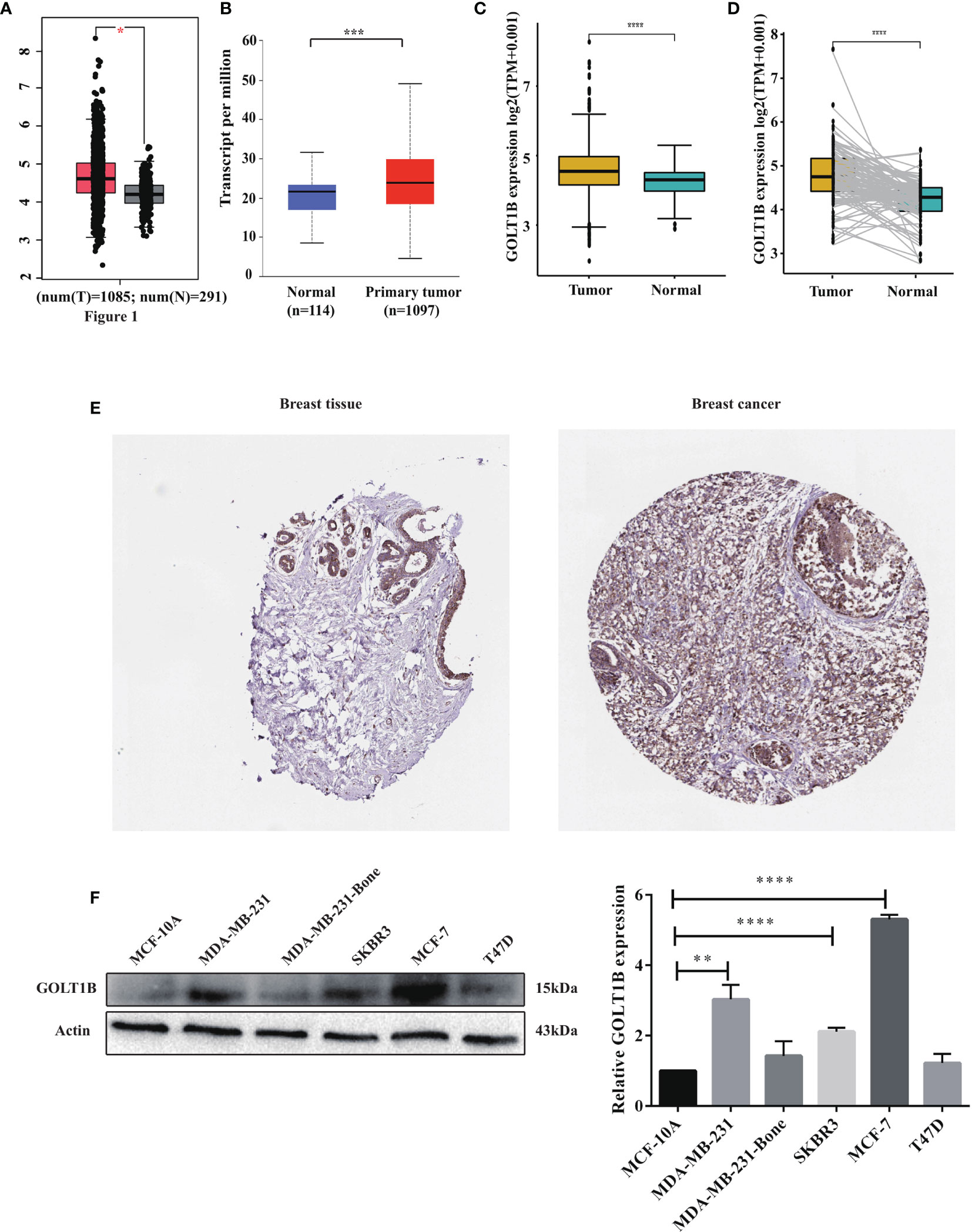
Frontiers | Multi-Omics Analyses Revealed GOLT1B as a ...
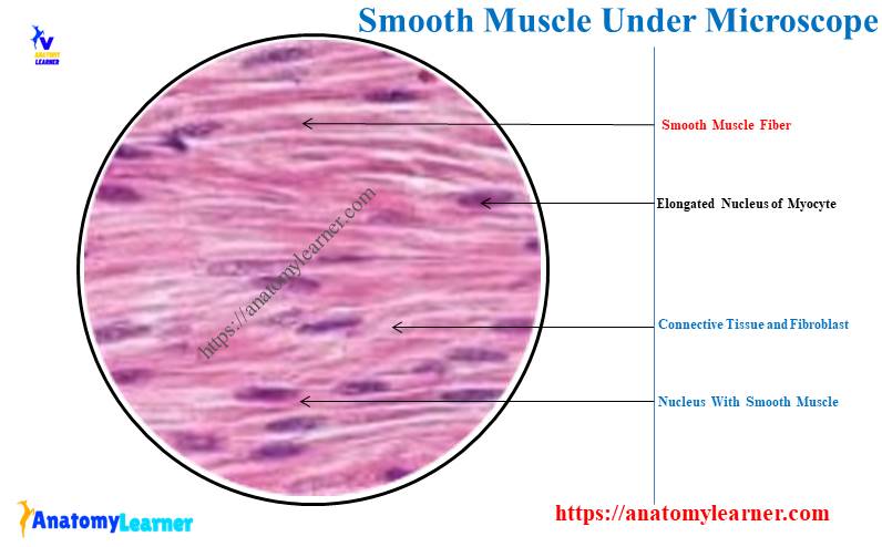
Smooth Muscle Under Microscope with Labeled Diagram ...

BIO - 168 Final Exam Study Guide Flashcards | Quizlet
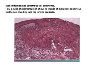
Oral epithelial tumor
A CLINICOPATHOLOGICAL EVALUATION OF NECK SECONDARIES IN ...
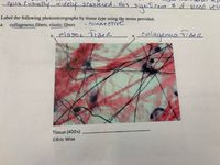
Answered: collagenous fibers, elastic fibers -… | bartleby

View Image
Solved Label the photomicrograph of thick skin. Stratum ...
Sebaceous (oil) gland • Hair follicle - ppt download
2,100+ Skin Histology Stock Photos, Pictures & Royalty-Free ...
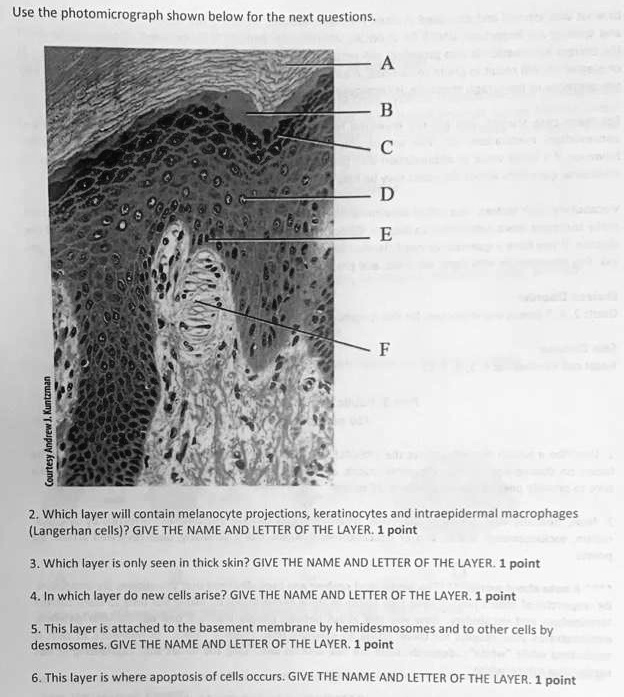
SOLVED: Use the photomicrograph shown below for the next ...

EXERCISE 4 QUIZ Flashcards | Quizlet

Localized delivery of immunotherapeutics: A rising trend in ...

Differentiation of Fibroblasts Into Myofibroblasts in the ...

Pin by Abby Kate on LAB FINAL | Thick skin, Langerhans cell ...
Respiratory System | histology
Animals | Free Full-Text | Naturally Produced Lovastatin ...
Integumentary System Overview
Frontiers | Cytochrome C as a potential clinical marker for ...

a) Photomicrograph showing islands of tumor cells arranged in ...

Regeneration and Assessment of the Endothelial Glycocalyx To ...
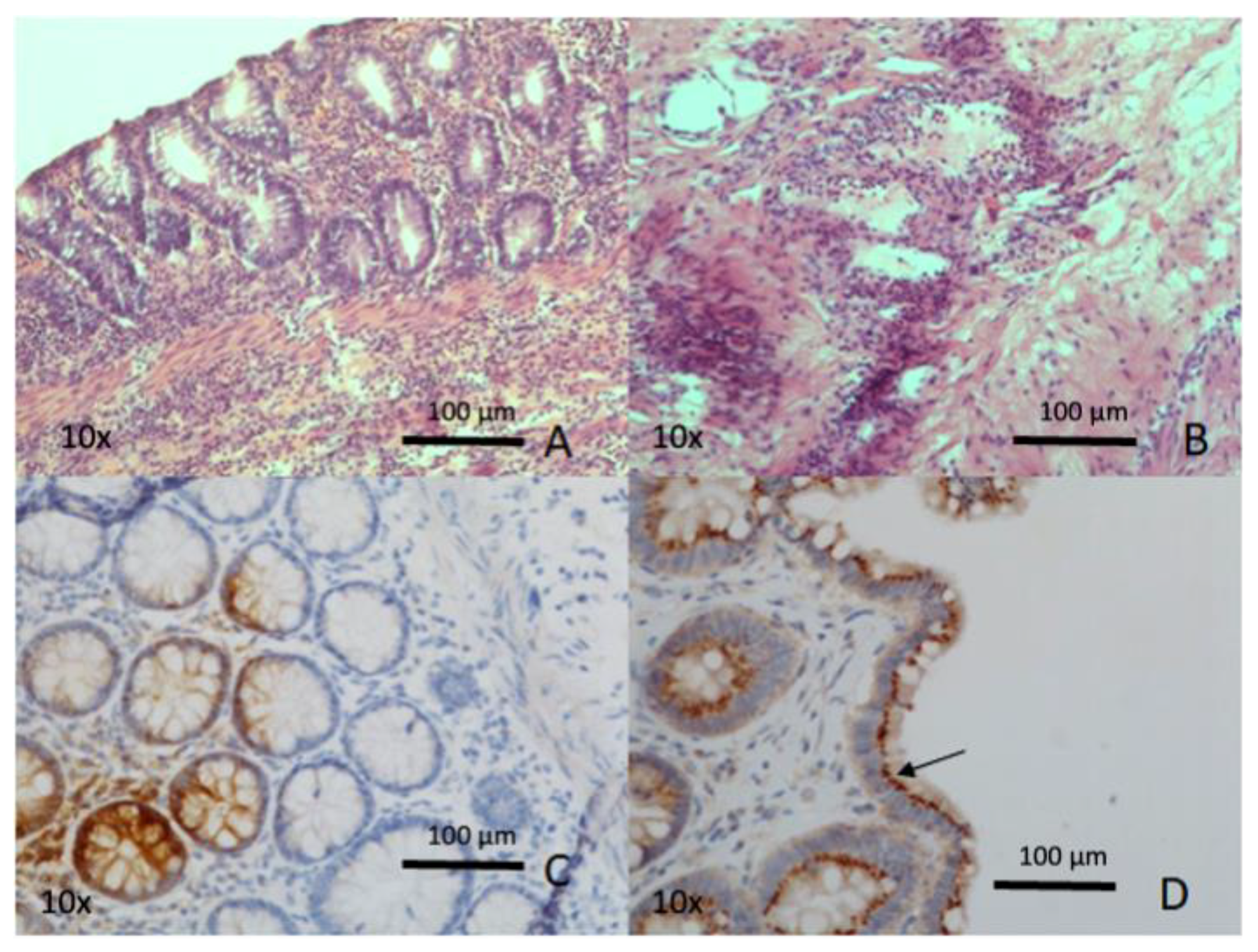
Biomedicines | Free Full-Text | Intestinal Ischemia: Unusual ...

Lab 9: Pre-Lab Homework Flashcards | Quizlet

Photomicrographs of a skin section showing the junction ...
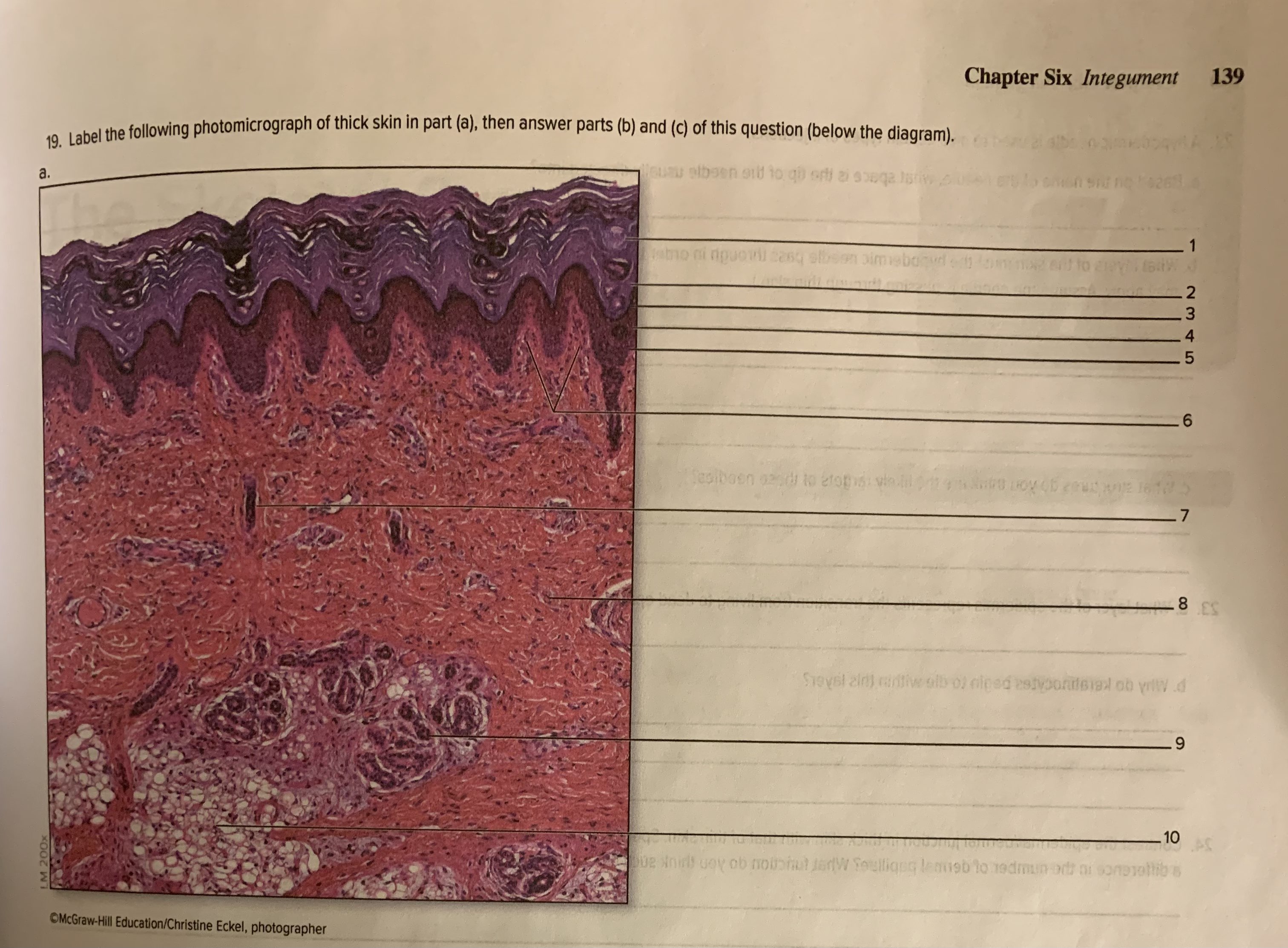
Solved Chapter Six Integument 139 19. Label the following ...
Stratified squamous non-keratinized epithelium
Chapter 5 Integumentary System. - ppt download

Identification of Nonepithelial Multipotent Cells in the ...

Epidermis | Biology for Majors II
Education Guide Special Stains and H & E Second Edition ...

photomicrograph of thick skin Diagram | Quizlet
Integumentary System Overview

Descending Dopaminergic Inputs to Reticulospinal Neurons ...
Phellogen hi-res stock photography and images - Alamy

222 Stratum Basale Images, Stock Photos & Vectors | Shutterstock
Immunofluorescent photomicrograph hi-res stock photography ...
Post a Comment for "40 label the photomicrograph of thick skin"